Conflicting Geometries
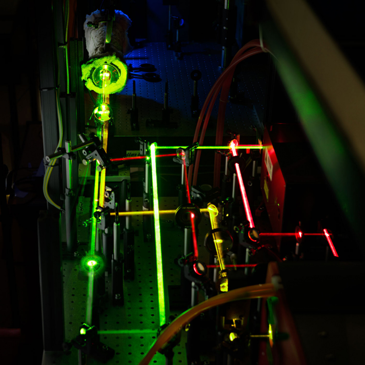
The challenge at the Centre for Sensor Systems (ZESS) Optics Lab was one of conflicting spatial demands: capturing the LED illumination array of the Fourier Ptychographic Microscopy (FPM) setup required a bottom-up perspective, while for the specimen and its technical mounting to be in the frame, top-down was necessary. To resolve this, we utilised the system’s integrated screen to monitor the specimen, allowing the camera to remain low to frame the hardware.

Shared Patience
Technical photography in a research context is a slow, deliberate method of working, but luckily it rarely is a solitary pursuit. Collaborating with Dr John Meshreki – a postdoctoral fellow at Professor Ivo Ihrke’s chair of Computational Sensorics and Communications Engineering at ZESS – once again highlighted the necessity of shared patience. He provided the necessary latitude to iterate through various lighting arrays and screen angles, ensuring the final frame reflected the precision of the research. Without the space to experiment with the lighting geometry, it would not have been possible to convey the nuances of the experimental setup.
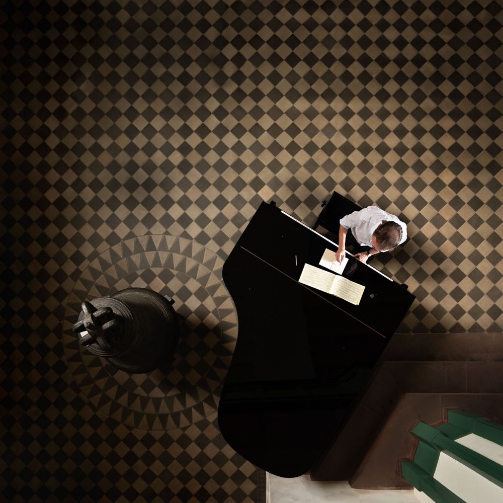
The Interdisciplinary Bridge
This experimental hardware is only one half of the equation; the second half is defined by mathematical reconstruction, for which John works closely with Prof Onofre Martorell, a mathematician from the University of the Balearic Islands. Complementing the lab photography with a documentation of a collaboration – demonstrating how physical sensorics and algorithmic theory converge – was a valuable opportunity. Onofre, working alongside Prof Michael Möller, applies machine learning and image processing to the raw data captured by the FPM setup. Their work, supported by the DFG research unit “Learning to Sense”, connects the darkroom with the realm of high-impact computational science, resulting in a super-resolution output that bridges the gap between raw data and diagnostic insight.
Technical Background
Fourier Ptychographic Microscopy represents a shift from chemical intervention to computational synthesis. Unlike traditional fluorescence microscopy – which often necessitates staining that can alter a specimen’s natural state – this technique reconstructs super-resolution images by capturing the phase of light through varied LED illumination angles. We used the intricate structures of a butterfly antenna to demonstrate this capability. By synthesising data from multiple green light sources, the system generates high-resolution results across a wide field of view. The resulting image is not a mere capture but a reconstruction of the microscopic universe in its unaltered form.
These images document the Fourier Ptychographic Microscopy (FPM) configuration within the optics laboratories at the Centre for Sensor Systems (ZESS). For the official research project details, scientific publications, and contact information for Prof Ivo Ihrke’s group, visit the Fourier Ptychography Microscopy Overview at ZESS, University of Siegen.